A Professional Guide for Medical Imaging Education and Clinical Practice
Fixed x ray machines remain a core imaging modality in hospital radiology departments due to their stable output, high image consistency, and adaptability to a wide range of clinical examinations. Understanding the technical parameters of fixed x ray systems is essential for medical students, radiology educators, and clinicians to ensure optimal image quality, radiation safety, and diagnostic accuracy.
This article provides a systematic explanation of key technical parameters used in modern fixed x ray machines, focusing on their physical principles and clinical relevance.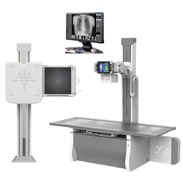
The generator power, expressed in kilowatts (kW), represents the maximum electrical power supplied to the X-ray tube.
Higher kW capacity allows simultaneous high kVp and mA output
Essential for:
Chest radiography
Bariatric patients
High-density anatomical regions (pelvis, lumbar spine)
30–50 kW: Basic radiography
65–80 kW: General hospital use
≥100 kW: Trauma centers and high-throughput departments
A higher kW rating improves exposure flexibility while maintaining short exposure times, reducing motion artifacts.
Kilovoltage peak (kVp) determines the maximum photon energy of the X-ray beam.
Controls penetration ability
Influences subject contrast
Affects patient radiation dose distribution
Chest X-ray: 100–125 kVp
Abdomen: 70–85 kVp
Extremities: 50–65 kVp
Higher kVp produces lower image contrast but improved penetration, suitable for thoracic imaging.
Tube current (mA) defines the number of electrons flowing through the X-ray tube per unit time, while exposure time (ms) controls the duration of exposure.
mAs = mA × exposure time
Determines total photon quantity
Directly proportional to image receptor exposure
Key factor in controlling image noise
High mA with short exposure time is preferred in chest and trauma imaging to minimize motion blur.
The focal spot is the area on the anode where electrons strike to produce X-rays.
Small focal spot (0.6 mm):
High spatial resolution
Fine bone and extremity imaging
Large focal spot (1.2 mm):
Higher heat capacity
General radiography
A smaller focal spot improves image sharpness but limits maximum tube loading.
Modern fixed x ray systems predominantly use digital flat-panel detectors (FPD).
Amorphous Silicon (a-Si) with Cesium Iodide (CsI)
Direct conversion (a-Se) detectors
Active area (e.g. 17″ × 17″)
Pixel size (typically 140–200 µm)
Detective Quantum Efficiency (DQE)
Higher DQE allows lower radiation dose while maintaining image quality.
AEC systems terminate exposure automatically when the detector receives sufficient radiation.
Reduces operator-dependent variability
Improves image consistency
Prevents under- or over-exposure
AEC is especially critical in chest and abdominal radiography where patient thickness varies significantly.
Anti-scatter grids are used to absorb scattered radiation before it reaches the detector.
8:1 – General radiography
10:1 or 12:1 – Chest and abdominal imaging
Higher grid ratios improve image contrast but require increased radiation dose.
Fixed x ray machines feature ceiling-mounted or floor-mounted tube supports with precise mechanical alignment.
Longitudinal and transverse tube travel
Tube-detector alignment accuracy
Vertical Bucky movement
Accurate positioning is critical for repeatable imaging and workflow efficiency.
Modern systems incorporate advanced image processing algorithms:
Dynamic range compression
Noise reduction
Edge enhancement
DICOM compatibility enables seamless integration with:
PACS
RIS
Hospital Information Systems (HIS)
Fixed x ray machines must comply with international standards such as:
IEC 60601-1
IEC 60601-2-54
FDA / CE regulations
Dose optimization strategies include:
Proper kVp selection
Use of AEC
High-efficiency detectors
A thorough understanding of fixed x ray machine technical parameters enables clinicians and educators to optimize image quality, ensure patient safety, and improve diagnostic confidence. Mastery of these parameters is essential for modern radiological practice and medical imaging education.
Other related articles:
Algeria: 25 Fixed X-Ray Machines Successfully Shipped & Delivered
How Hospitals Define Technical Specifications for Fixed X Ray Machines
How Hospitals Choose X-Ray Machine Suppliers